March 16, 2015
Maintaining 3D body shape through tension
IST Austria Professor Heisenberg and collaborators unravel new mechanism for maintenance of 3D body shape in current issue of Nature • YAP gene controls tissue tension needed to resist gravity • Breakdown of actomyosin network reduces tension

How does a vertebrate organism develop its shape? It all starts in the developing embryo: individual cells that divide, grow and mature progressively arrange themselves into distinct tissues and organs, which together form a three-dimensional living body. Tissue tension is important for the development of body shape as the embryo is exposed to gravity and needs to withstand these external forces. Tissue tension is generated by cortical tensile forces transmitted via cell-cell contacts, or via adhesions between cells and the extracellular environment. But what controls this tissue tension? It looks like it’s all down to a gene called YAP, which influences both the mechanical forces acting from within cells and on the extracellular environment. This was discovered in a multi-institutional research project spearheaded by IST Austria Professor Carl-Philipp Heisenberg’s group, Hiroshi Nishina’s team at the Medical and Dental University in Tokyo, Japan, and Makoto Furutani-Seiki’s lab from the University of Bath in the UK,. The results are published in a Letter in Nature on March 16.
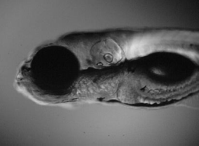
How did the scientists make this discovery? It became evident that YAP played an important role in the maintenance of body shape when they screened many genetic mutants of zebrafish and medaka fish embryos, both model organisms used to study vertebrate development. In mutants where YAP is altered such that it no longer functions, the overall tension in the developing tissue is reduced. As a result, the 3D embryo succumbs to gravity and the body flattens.
Why is that the case? The major machinery generating cellular force through contraction is actomyosin, which is a complex of myosin motor proteins interacting with cytoskeletal structures such as actin fibres in the cell cortex, just underneath the cell surface. If the formation of this cortical actomyosin network is disrupted, as is the case in the YAP mutants, the cell lacks a proper structural scaffold and cortical tension is reduced. However, that is not the whole story. Actomyosin contraction also stimulates the assembly of fibronectin in the extracellular matrix. The extracellular matrix is a collection of molecules that help the cells stick together and fibronectin is one of those molecules. In the absence of YAP and actomyosin contraction, fibronectin fibrils are not adequately produced and hence the extracellular matrix is not properly organised. The result: dividing cells no longer stack properly on top of one another and tissue becomes misaligned.
That YAP is required to maintain tissue tension is a hitherto unknown function of this gene. It was previously known to act as a mechanosensor of cellular environment stiffness, thereby regulating cell proliferation. This new data indicates that YAP also acts as a mechanoregulator. YAP is a gene present across the animal kingdom and the team of scientists could show that YAP function in the maintenance of 3D tissue shape is conserved in human cells. These findings could be useful in the context of regenerative medicine. It could help understand how complex organs are organized by force-mediated tissue alignment.


