December 11, 2024
The Distinct Nerve Wiring of Human Memory
ISTA biologists team up with neurosurgeons to unravel the human brain’s specificities
The black box of the human brain is starting to crack open. Although animal models are instrumental in shaping our understanding of the mammalian brain, scarce human data is uncovering important specificities. In a paper published in Cell, a team led by the Jonas group at the Institute of Science and Technology Austria (ISTA) and neurosurgeons from the Medical University of Vienna shed light on the human hippocampal CA3 region, central for memory storage.
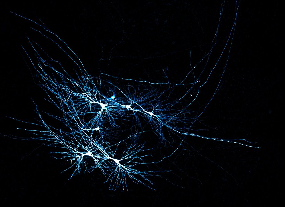
Many of us have relished those stolen moments with a grandparent by the fireplace, our hearts racing to the intrigues of their stories from good old times, recounted with vivid imagery and a pinch of fantasy. Our human brain has a remarkable capacity for storing and recollecting memories over a lifetime. A physical space, a smell, or a familiar situation can alone bring back a memory, and our brain uses these associations to complete the pattern. Although the human brain is optimized for this purpose, we are only starting to understand how it integrates information about our surroundings. This pattern-completion process is a remarkable computational property of our brain called associative memory.
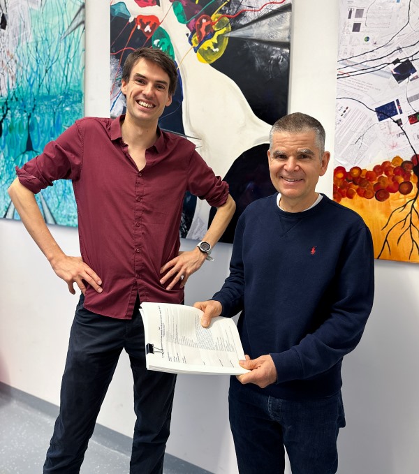
The bulk of our neuroscience knowledge about the brain stems from well-studied animal models, like rodents, which are indispensable for science. But is the human brain simply a scaled-up version of the mouse brain, or does it have distinct features that make it human? Now, researchers at the Institute of Science and Technology Austria (ISTA) and neurosurgeons at the Medical University of Vienna shed light on how the human brain forms and retrieves associative memories. Magdalena Walz Professor for Life Sciences at ISTA Peter Jonas and ISTA postdoc Jake Watson, who initiated the collaboration with Professor Karl Rössler from the Department of Neurosurgery of the Medical University of Vienna, examined samples from epilepsy patients who underwent neurosurgery to gain insights directly from intact, living human tissue.
Distinct human features of memory formation
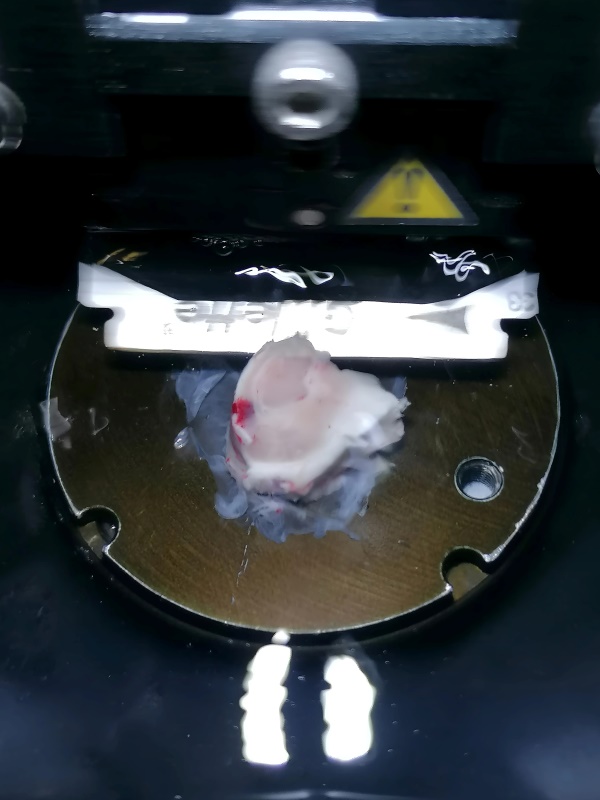
The brain’s center for learning and associative memory is the hippocampus. Within it, a region called CA3 is responsible for storing and processing information and completing patterns. Because healthy human material is scarce, most studies have so far focused on animal models. Jonas and Watson overcame this problem by teaming up with Rössler, a neurosurgeon specializing in treatment-resistant forms of epilepsy. “While patients undergoing neurosurgery have a wide variety of clinical presentations, Prof. Rössler identified a subpopulation of epilepsy patients who presented an intact hippocampus,” says Jonas. The scientists could not possibly miss this opportunity. “In this form of epilepsy, a unilateral resection of the hippocampus is necessary to ensure the patients have a chance to recover and lead an epilepsy-free life,” explains Jonas. Thus, the team could obtain intact hippocampal tissue from 17 epilepsy patients with informed consent.
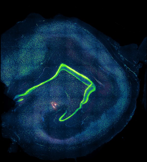
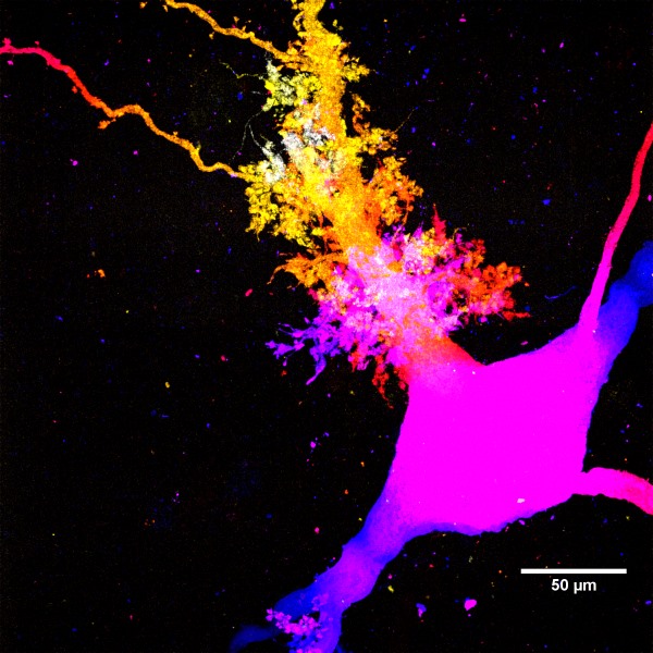
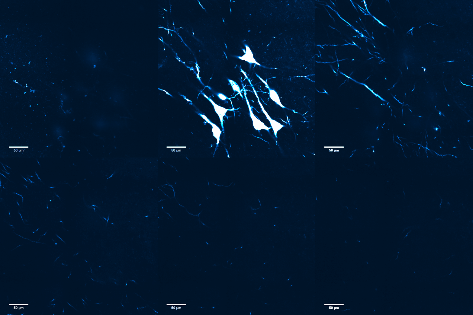
The researchers combined cutting-edge experimental techniques—multicellular patch-clamp recording to measure dynamic functional properties of neurons and super-resolution microscopy—with modeling and made eye-opening findings. Far from being a scaled-up version of the well-studied mouse hippocampus, the neural connectivity in the human CA3 region was sparser, and its synapses—the connections that allow signals to be passed between the neurons—appeared more reliable and precise. Thus, the team uncovered distinct properties of the human brain’s wiring.
“It felt like we didn’t know a thing”
Despite the important differences in the cellular structure and synaptic connectivity of the human hippocampus compared to that of mice and rats, data from animal models remains very important. It serves as a reference and helps scientists develop the technology for studying human tissue. “Coming from a background working with rodents, it can feel like everything about the hippocampus is already known,” says Watson. “As soon as I started examining the first patient samples, I realized how much we didn’t know about the human hippocampus. Although this is the best-studied brain region in rodents, it felt like we didn’t know a thing about human physiology, cellular organization, or connectivity.” Thus, based on their experience working with rodent hippocampal tissue, Watson and Jonas needed to find new ways to re-examine this part of the brain in humans.
Modeling the human brain’s computational power
With their experimental data, the team sought to build a model of the computational power of the CA3 network in the human hippocampus. They realized the human-specific circuitry and synaptic connectivity allowed them to measure the extent to which memories were stored and retrieved reliably. “We could test how many patterns fit in this model. This helped us demonstrate that the human-specific sparse synaptic connectivity and enhanced synaptic reliability increased storage capacity,” says Jonas. In other words, they uncovered how the human CA3 network codes information efficiently to maximize associations and memory storage.
The best day in a physiologist’s career
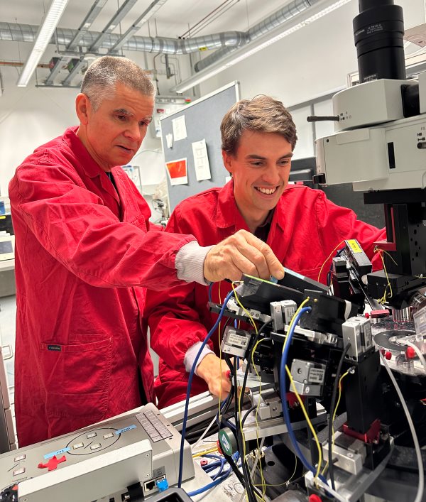
The present study helps change how scientists and healthcare professionals perceive the human brain. “Our work highlights the need to rethink our understanding of the brain from a human perspective. Future research on brain circuitry, even if using rodent model organisms, must be conducted with the human brain in mind,” says Jonas. According to the scientists, this work is the fruit of a synergy between the right neurosurgeon and the right physiologists. “Prof. Rössler is very keen on promoting basic research and has developed sophisticated techniques to extract patient tissue in the best possible condition for laboratory examination,” emphasizes Watson. This collaboration has given the ISTA researchers access to a scarce resource in science: intact, living human brain tissue. Since tissue availability depended on the surgeries, the scientists only received new biological material sporadically every few months. This has impacted their lab’s logistics: they often needed to interrupt all projects using non-human material on short notice and clear the space to receive and examine the fresh human samples. “It felt surreal thinking that the epilepsy patient who underwent neurosurgery the same morning was recovering in hospital while we were examining an intact and living slice of tissue from their brain,” says Watson. “Looking back, the best day in my career as a physiologist was when the first human tissues arrived in our lab.”
Publication:
Jake F. Watson, Victor Vargas-Barroso, Rebecca J. Morse-Mora, Andrea Navas-Olive, Mojtaba R. Tavakoli, Johann G. Danzl, Matthias Tomschik, Karl Rössler, and Peter Jonas. 2024. Human hippocampal CA3 uses specific functional connectivity rules for efficient associative memory. Cell. DOI: 10.1016/j.cell.2024.11.022
Funding information:
This project was supported by funding from the European Research Council (ERC) under the European Union’s Horizon 2020 research and innovation program (Advanced Grant no. 692692; Marie Skłodowska-Curie Actions Individual Fellowship no. 101026635), the Austrian Science Fund (FWF; grant PAT 4178023; grant DK W1232), the Austrian Academy of Sciences (DOC fellowship 26137), and a NOMIS-ISTA fellowship.
Information on human patient tissue samples:
Human tissue samples were obtained with informed patient consent from 17 individuals with temporal lobe epilepsy. This work was approved by the Ethics Committee of the Medical University Vienna (MUW) (EK Nr: 2271/2021). Further information can be found in the paper’s experimental model and study participant details section.
Information on human postmortem tissue samples:
Three blocks (each approximately 1 cm3) of postmortem tissue were acquired from the Normal Ageing Brain Collection Amsterdam (NABCA) biobank (Project agreement METC: 2023.0733; ISTA Ethics committee application: 2023-03). Further information can be found in the paper’s experimental model and study participant details section.
Information on animal studies:
To better understand fundamental processes, for example, in the fields of neuroscience, immunology, or genetics, the use of animals in research is indispensable. No other methods, such as in silico models, can serve as an alternative. The animals are raised, kept, and treated according to strict regulations. The research with animals was conducted at ISTA.


