June 11, 2018
Two bilateral French-Austrian research projects start at IST Austria
Research on cell division and synapse function funded by FWF and ANR - Project leaders at IST Austria are Carl-Philipp Heisenberg und Johann Danzl
Two joint projects between labs at the Institute of Science and Technology Austria and at French research institutes, funded by the Austrian Science Fund FWF and the Agence Nationale de la Recherche ANR, are kicking-off. The lab of Carl-Philipp Heisenberg at IST Austria, together with the lab of Alex MacDougall at Centre national de la recherche scientifique (CNRS), will study how polarity, shape and mechanics of cells control cell division. Johann Danzl at IST Austria partners with the lab of Olivier Thoumine at the Interdisciplinary Institute for Neuroscience in Bourdeaux to study which role synaptic adhesion molecules play in synapse function, using optically controlled molecules and high-resolution optical imaging. The bilateral French-Austrian Joint Research Projects are funded for three years with a total of around 250,000€ each.
What are the rules controlling cell division?
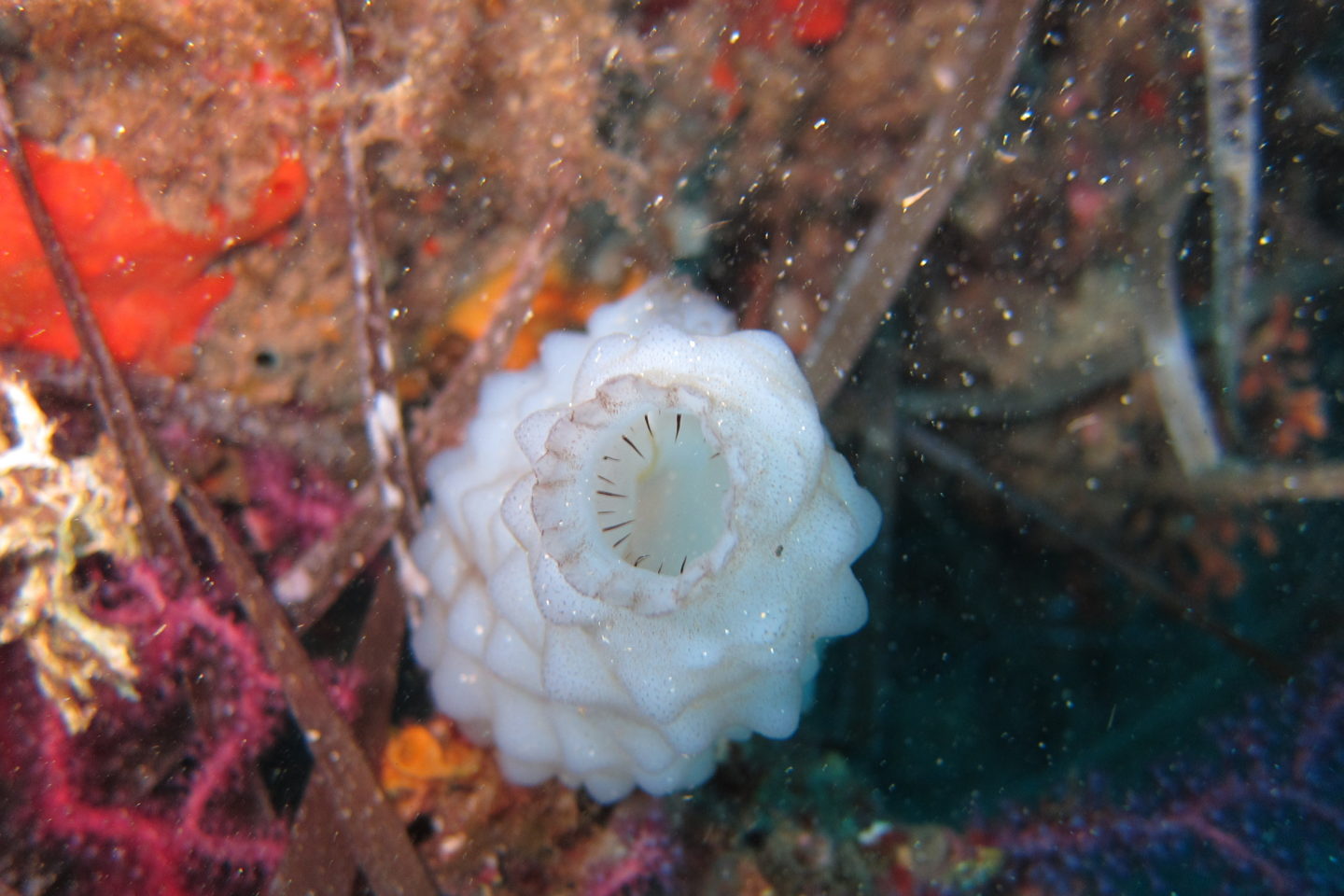
Ascidians, or sea squirts, are closely related to vertebrates but look rather unremarkable. Their name derives from the Greek word for “little bag”, and indeed the 1mm to 10 cm long sea animals resemble odd blobs. While they look unassuming as adults, the embryos have a remarkable characteristic: they consist only of a small number of cells, and the positioning and timing of cell divisions are identical between different individuals of the same species – and even between species. But what are the rules that govern this so-called “invariate cleavage pattern”? In the joint project, the Heisenberg and MacDougall labs will investigate this question in the ascidian Phallusia mammillata.
Maternal factors and gene-regulatory networks are known to affect cell division and position in the ascidian embryo. However, cells in the embryo do not exist in isolation, but press against each other. Cues are likely to spread between cells through adhesion, which transmits mechanical forces across cells. But how do these physical forces influence cleavage pattern and cell position? The Heisenberg and MacDougall labs will pool their expertise to answer this question.
The McDougall lab previously showed that the ascidian cell division pattern depends on the positioning of the so-called spindle along the cell axis. The spindle is the structure inside a dividing cell that distributes copies of the cell’s genetic material between the new cells, and its position along the so-called apicobasal cell axis influences the position of division. In the new project, the labs will combine molecular, cellular and biophysical experiments to look at how apicobasal cell polarization interacts with cell shape to orient cell division and give shape to the embryo. This project combines the expertise of the ascidian laboratory led by MacDougall with the expertise of the Heisenberg laboratory in measuring the mechanical properties of cells to unravel the complex interplay between apicobasal polarity and cell shape.
Which role do synaptic adhesion molecules play in synaptic connections?
Signals in our brains are sent from one neuron to another via their connections, the synapses. The message itself is sent through chemicals called neurotransmitters, which are released by the pre-synaptic neuron and sensed through receptors on the post-synaptic neuron. But the pre- and post-synaptic neurons are also structurally connected through so-called adhesion molecules. These neuronal adhesion molecules, such as neurexins on the pre-synaptic neuron and neuroligins on the post-synaptic neuron, play important roles in wiring, sculpting and maintaining synaptic connections. But how do synaptic adhesion molecules control the formation of synapses? Johann Danzl at IST Austria and Olivier Thoumine investigate this question by putting the adhesion molecules under light control, helping to understand synaptic development and function.

© Johann Danzl/IST Austria
In the project, Danzl and Thoumine will use optogenetically controlled synaptic adhesion molecules, which can be switched on and off with light at exactly defined time points. In this way, the researchers can follow the formation of synapses in living neurons as adhesion molecules are switched from the off into the on state. The Thoumine lab is specialized in neuronal adhesion proteins, with expertise in single molecule imaging, computation and electrophysiology to study adhesion molecules and their dynamics at the single-molecule level. The lab of Johann Danzl at IST Austria has expertise in imaging using super-resolution nanoscopy, which has a much higher resolution than conventional light microscopy, and optical control of photoswitchable molecules. This allows them to image the fine structural features of neuronal cells and synapses. Bringing the expertise of these labs together, the project will enable the scientists to dynamically and quantitatively describe and regulate adhesion protein clustering and function at synapses.


