June 17, 2021
Developing new technologies to visualize the inner workings of cells
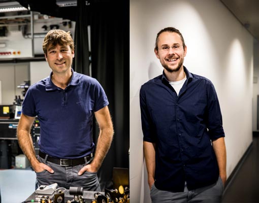
IST Austria professors Johann Danzl and Florian Schur receive a grant from the Chan Zuckerberg Initiative
Imaging molecules, cells, and tissues is central to fundamental biological and biomedical research, allowing scientists to better understand biological mechanisms in health and disease. The Chan Zuckerberg Initiative (CZI) aims to move biology and medicine forward by supporting researchers in developing cutting-edge imaging technologies and to share them with the research community. Two IST Austria professors, Johann Danzl and Florian Schur, are grantees of CZI’s recent “Visual Proteomics” call to advance technology to observe the inner workings of cells at near-atomic resolution.
Within their joint project, Florian Schur and Hans Danzl will develop combined light and electron microscopy to visualize biological samples in a near-native state at low temperatures with extremely high precision. Light and electron microscopy are two highly complementary imaging modalities. The technology developed by the scientists will identify specific individual proteins in cells with nanometer-scale resolving low temperature (cryo) light microscopy, will unravel the structural context of these molecules, and ultimately determine their 3D structure inside their natural cellular environment with electron microscopy. If successful, the project will unleash the full synergistic potential of light and electron microscopy by allowing nanometer-precise correlation between them. Ultimately, this new technology will be shared with the scientific community to enable researchers to gain insight into biological and biomedical mechanisms by revealing molecular machineries in the most native state.
The grant to Florian Schur and Hans Danzl follows the recent CZI funding of Robert Hauschild, a staff scientist at IST Austria who received a CZI Imaging Scientist program grant in December 2020. Robert Hauschild’s project will develop tools that help other researchers utilize their microscopes to the fullest extent. He is working on hardware for sample manipulation and environmental control, among other purposes, and on automation software. He also supports the work of the imaging core facility staff with accessories and protocols to evaluate and maintain microscope performance. His aim is to make all his tools available as open source in a way they can be implemented by non-specialists.
