September 23, 2020
Feeling Like a Cell
A researcher investigates how immune cells feel their way through our bodies.

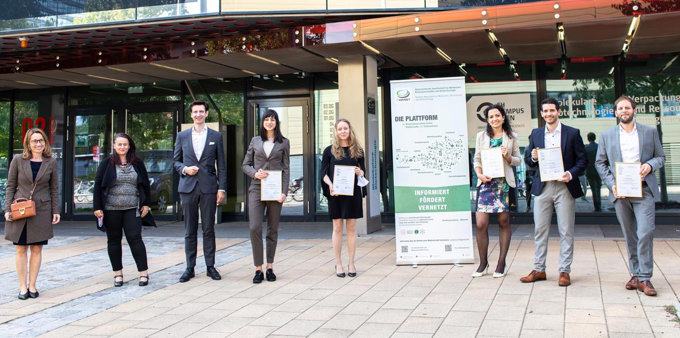
Every second countless cells move through our bodies looking for something to eat. In her PhD thesis, Aglaja Kopf from the Sixt research group at the Institute of Science and Technology Austria (IST Austria) investigated how they feel their way through the tissue. For this, she now received the PhD Award from the Austrian Association of Molecular Life Sciences and Biotechnology (ÖGMBT).
White blood cells—the leukocytes—are on the hunt. They are part of our bodies’ immune system moving quickly around and looking for intruders to eat. By doing this, they fight off infections and diseases. But how exactly they find their way through a complex meshwork of connective tissues and cells is still a matter of research. Aglaja Kopf from the Sixt research group at the Institute of Science and Technology Austria (IST Austria) found a new keystone in our understanding of leukocyte movement and how these extremely fast moving cells sense and maintain their own shape. For her PhD thesis, she now received the PhD Award of the Austrian Association of Molecular Life Sciences and Biotechnology (ÖGMBT) on September 21, 2020.
On their journey through the body, leukocytes face a myriad of obstacles, forcing these cells to closely sample their surrounding for optimal migration routes. They do so by stretching multiple exploratory protrusions into their immediate vicinity that, whenever the cell has decided for a specific route, need to be retracted in a coordinated fashion. Otherwise, the cell would become entangled or in the worst case rupture and die. However, how fast and branched out cells are able to sense and integrate their own shape is still poorly understood.
Inside the Cells

In her thesis Aglaja Kopf investigated the role of microtubules in the leukocytes’ sensing of their own shape. Microtubules are part of the cytoskeleton, the strands of protein in a cell that are essential for cell division and allow the internal transport of molecules and information. They are hollow cylinders made of proteins with a diameter of 20 to 30 nanometers (a billionth of a meter) that radiate outwards from a microtubule-organizing center in the cell towards its outer membrane. On their ends they continuously grow and break down, forming filaments of variable lengths. But how do microtubules contribute to the shape sensing of motile cells?
First, the microtubule-organizing center acts as a pathfinder by moving into the winner protrusion, the arm along which the cell will move. This is followed by a local break down of microtubule filaments in the looser protrusions which soon after are retracted. One explanation of why microtubules start disappearing once the organizing center has moved into a specific protrusion is, that these cylinders are rigid and grow in straight trajectories. So when the cell moves its organizing center around a corner or encounters an obstacle, the microtubules fracture releasing signal molecules that steer the movement of the cell away. This allows it to pass through tight constrictions in tissues when hunting for intruding cells and viruses.
When microtubules were destroyed in migrating leukocytes in an experiment, the cells became entangled because exploratory protrusions kept on growing, making it impossible for the cell to decide for a specific migration route, which frequently resulted in fragmentation and ultimately cell death.
Wandering Cancer and Sensing Neurons
Investigating the sensing of cells is not only important in leukocytes, but also may be relevant for cancer growth and the formation of neurons. Cancer is formed by abnormally migrating cells, so this newly discovered mechanism may help to understand how cancer cells can form metastases in distant parts of the body, and how to stop them from doing so.
Neurons in the brain first grow many connections to other neurons but then maintain only certain ones. The first indication of degradation in the not needed arms is the disassembly of microtubules. The here discovered mechanism also contributes to this process, but how exactly it plays into neuronal circuit formation is still an open question.
In her research Aglaja Kopf used advanced 3D microscopy to film and study the movement of leukocytes both in fabricated micro-channels and in model organisms. She took bone marrow cells, turned them into immune cells, and activated them by introducing cell wall fragments from E. coli bacteria. This worked like a lure for a predator making the leukocytes become highly motile.
All cells in our bodies have some kind of sensing capabilities for their own shape. Studying these could allow us to treat diseases better and support our immune cells on their hunt inside our bodies.
Publication
Kopf, A. 2019. The implication of cytoskeletal dynamics during leukocyte migration. IST Austria. https://doi.org/10.15479/AT:ISTA:6891
Funding information
The IST Austria project part was supported by funding from FWF, grant number W01250-B20, Nano-Analytics of Cellular Systems.


