June 10, 2022
How to Make Cells Glow
The mechanisms behind green fluorescent proteins. Image © Kasumi Kishi, ISTA
Would you like to have green glowing eyes? While we embrace diversity of all kinds, such genetic modification could raise some eyebrows. Yet, it is possible to make cells glow green in living organisms, in fact, it has become a commonly used method in cell biology. But how does it work and what is it used for?
The dog’s name was Ruppy and he was glowing in the dark. This experiment by the Seoul National University in South Korea seems like a debatable proof-of-concept, the method of making living cells glow however has revolutionized biological research. Green fluorescent proteins, GFPs in short, emit green light when exposed to blue or ultraviolet one. The high energy of the blue and ultraviolet light adds energy to the molecules of the protein. A moment later, they release that energy again but at a different wavelength – with GFPs this is 500 to 570 nanometers, which we humans perceive as green light.
“I help the Institute’s scientists to genetically modify all kinds of cells to carry the fluorescent proteins,” says Mark Smyth, virus service technician in the Molecular Biology Service at the Institute of Science and Technology Austria (ISTA). For this, he designs special viruses that sneak the genetic code for the GFP into the targeted cells, usually alongside the code of a particular protein of interest. “The infected cells then produce the GFP and, if included, the protein of interest. The GFP can be made separately to the protein of interest, or they both can be made as one long protein. The glowing GFP acts then like a little beacon that can be tracked by a microscope,” Smyth lays out the procedure. When the desired protein is fused with GFP, the scientists can even identify exact location of the protein within the cell. GFP fluorescence can also be used to check whether a protein was successfully introduced into the cell and to track how cells move around and how they change over time.
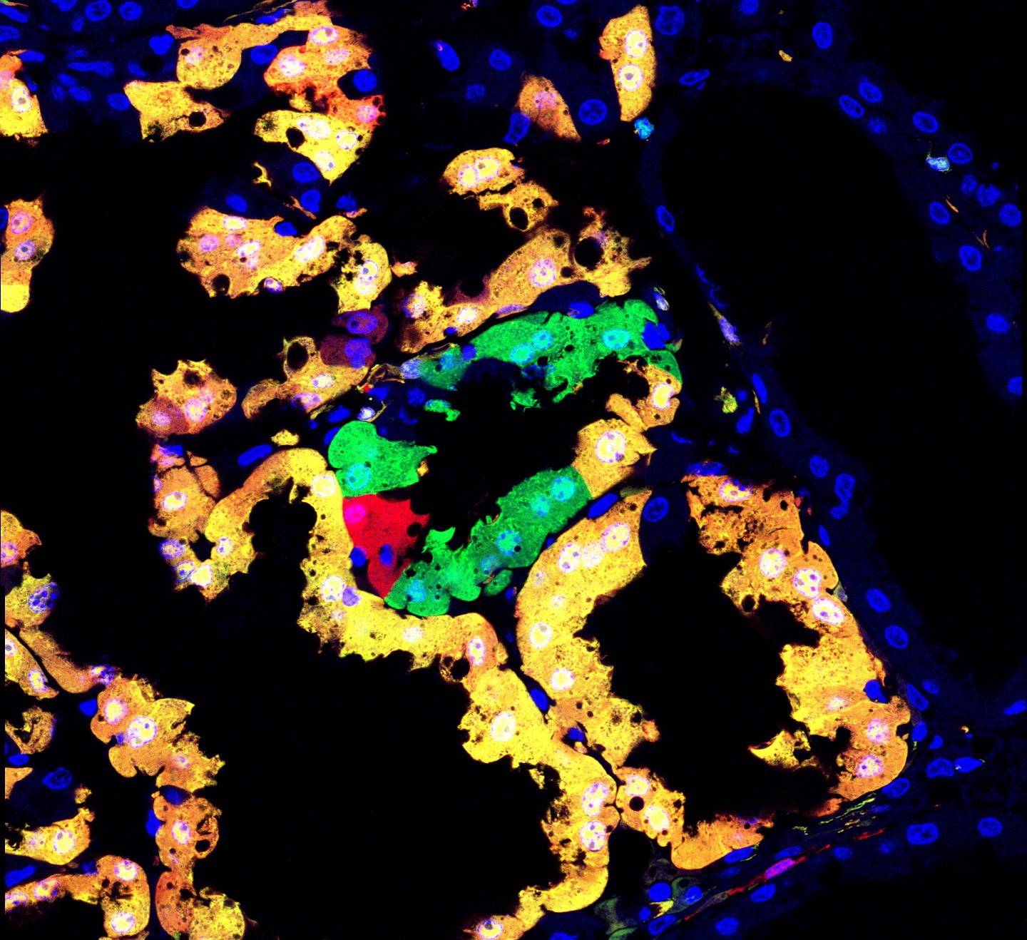
Cells in the mammary gland (left) and in the heart (right) of a mouse. The Hippenmeyer group at ISTA does not only use green, but various fluorescent proteins for their research. © Nicole Amberg / ISTA; Simon Hippenmeyer / ISTA
Enlightenment used by Science
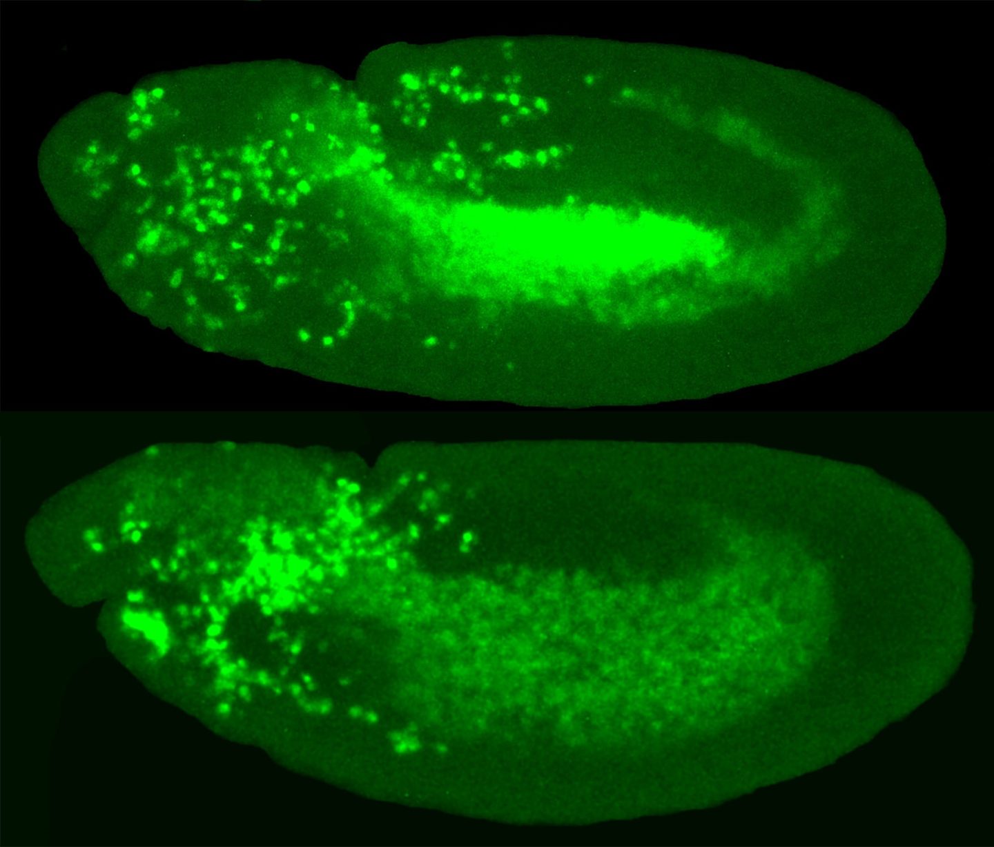
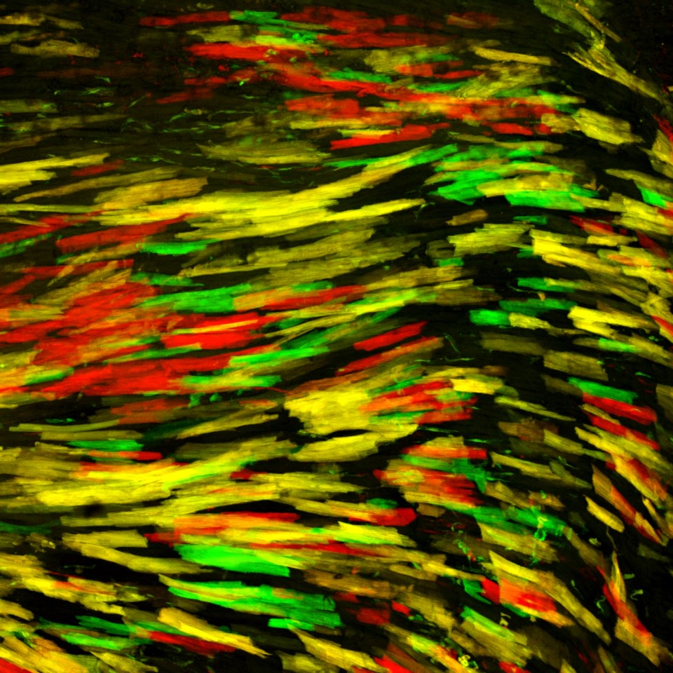
At ISTA, this tool has led to plenty of insights across biological disciplines: Researchers around Daria Siekhaus have applied it to fruit flies to understand how immune cells enter new tissue, the group of Carl-Philipp Heisenberg used it to investigate early embryonal development in zebrafish, Jiří Friml and his team gained deeper knowledge about plant growth by making seedlings glow, and Simon Hippenmeyer’s group developed a novel method for mouse genetics, where genetic mutations in individual cells can be labeled and tracked with such fluorescent colors. Michael Sixt’s lab has used it to study how the internal skeleton of immune cell moves and rearranges itself, specifically how these changes allow the cell to move from one place to another.
Interestingly, the green fluorescent proteins are no crazy laboratory invention. They were discovered in nature, in corals and other marine organisms. Researchers first isolated them sixty years ago from the gracefully glowing jellyfish Aequorea victoria and received the 2008 Nobel Prize for it. Here, understanding the beauty of natural life had led once again to a plethora of technological and scientific breakthroughs.
