January 9, 2024
Stranger than Friction: A Force Initiating Life
Scientists examine how friction forces propel development in a marine organism
As the potter works the spinning wheel, the friction between their hands and the soft clay helps them shape it into all kinds of forms and creations. In a fascinating parallel, sea squirt oocytes (immature egg cells) harness friction within various compartments in their interior to undergo developmental changes after conception. A study from the Heisenberg group at the Institute of Science and Technology Austria (ISTA), published in Nature Physics, now describes how this works.
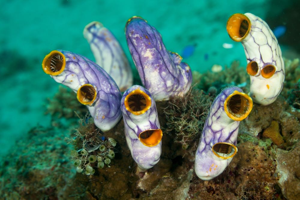
The sea is full of fascinating life forms. From algae and colorful fish to marine snails and sea squirts, a completely different world reveals itself underwater. Sea squirts or ascidians in particular are very unusual: after a free-moving larvae stage, the larva settles down, attaches to solid surfaces like rocks or corals, and develops tubes (siphons), their defining feature. Although they look like rubbery blobs as adults, they are the most closely related invertebrate relatives to humans. Especially at the larval stages, sea squirts are surprisingly similar to us.
Therefore, ascidians are often used as model organisms to study the early embryonic development of vertebrates to which humans belong. “While ascidians exhibit the basic developmental and morphological features of vertebrates, they also have the cellular and genomic simplicity typical of invertebrates,” explains Carl-Philipp Heisenberg, Professor at the Institute of Science and Technology Austria (ISTA). “Especially the ascidian larva is an ideal model for understanding early vertebrate development.”
His research group’s latest work, published in Nature Physics, now gives new insights into their development. The findings suggest that upon fertilization of ascidian oocytes, friction forces play a crucial role in reshaping and reorganizing their insides, heralding the next steps in their developmental cascade.
Decoding oocyte transformation
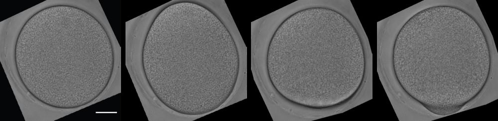
Oocytes are female germ cells involved in reproduction. After successful fertilization with male sperm, animal oocytes typically undergo cytoplasmic reorganization, altering their cellular contents and components. This process establishes the blueprint for the embryo’s subsequent development. In ascidians, for instance, this reshuffling leads to the formation of a bell-like protrusion—a little bump or nose shape—known as the contraction pole (CP), where essential materials gather that facilitate the embryo’s maturation. The underlying mechanism driving this process, however, has been unknown.
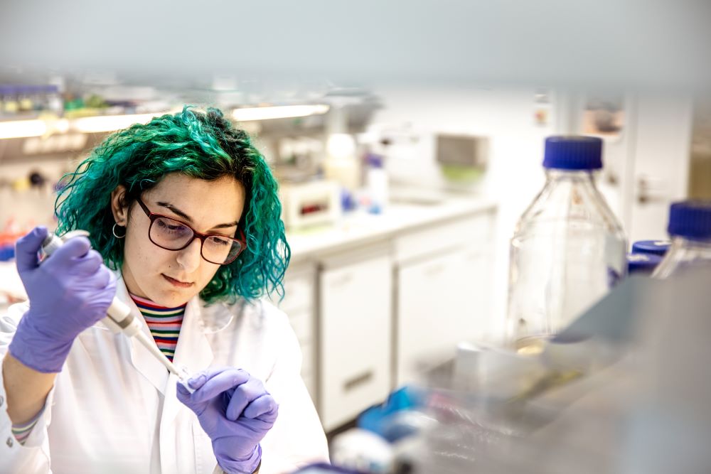
A group of scientists from ISTA, Université de Paris Cité, CNRS, King’s College London, and Sorbonne Université set out to decipher that mystery. For this endeavor, the Heisenberg group imported adult ascidians from the Roscoff Marine Station in France. Almost all sea squirts are hermaphrodites, as they produce both male and female germ cells. “In the lab, we keep them in saltwater tanks in a species-appropriate manner to obtain eggs and sperm for studying their early embryonic development,” says Silvia Caballero-Mancebo, the first author of this study and previous PhD student in the Heisenberg lab.
The scientists microscopically analyzed fertilized ascidian oocytes and realized that they were following very reproducible changes in cell shape leading up to the formation of the contraction pole. The researchers’ first investigation focused on the actomyosin (cell) cortex—a dynamic structure found beneath the cell membrane in animal cells. Composed of actin filaments and motor proteins, it generally acts as a driver for shape changes in cells.
“We uncovered that when cells are fertilized, increased tension in the actomyosin cortex causes it to contract, leading to its movement (flow), resulting in the initial changes of the cell’s shape,” Caballero-Mancebo continues. The actomyosin flows, however, stopped during the expansion of the contraction pole, suggesting that there are additional players responsible for the bump.
Friction forces impact cell reshaping
The scientists took a closer look at other cellular components that might play a role in the expansion of the contraction pole. In doing so, they came across the myoplasm, a layer composed of intracellular organelles and molecules (related forms of which are found in many vertebrate and invertebrate eggs), positioned in the lower region of the ascidian egg cell. “This specific layer behaves like a stretchy solid—it changes its shape along with the oocyte during fertilization,” Caballero-Mancebo explains.
During the actomyosin cortex flow, the myoplasm folds and forms many buckles due to the friction forces established between the two components. As actomyosin movement stops, the friction forces also disappear. “This cessation eventually leads to the expansion of the contraction pole as the multiple myoplasm buckles resolve into the well-defined bell-like-shaped bump,” Caballero-Mancebo adds.
The study provides novel insight into how mechanical forces determine cell and organismal shape. It shows that friction forces are pivotal for shaping and forming an evolving organism. However, scientists are only at the beginning of understanding the specific role of friction in embryonic development. Heisenberg adds: “The myoplasm is also very intriguing, as it is involved in other embryonic processes of ascidians as well. Exploring its unusual material properties and grasping how they play a role in shaping sea squirts, will be highly interesting.”

Publication:
S. Caballero-Mancebo, R. Shinde, M. Bolger-Munro, M. Peruzzo, G. Szep, I. Steccari, D. Labrousse-Arias, V. Zheden, J. Merrin, A. Callan-Jones, R. Voituriez & C.P. Heisenberg. 2024. Friction forces determine cytoplasmic reorganization and shape changes of ascidian oocytes upon fertilization. Nature Physics. DOI: 10.1038/s41567-023-02302-1
Funding information:
This project was supported work was supported by a Joint Project Grant from the FWF (I 3601-B27).
Information on animal studies:
In order to better understand fundamental processes, for example, in the fields of neuroscience, immunology, or genetics, the use of animals in research is indispensable. No other methods, such as in silico models, can serve as alternative. The animals are raised, kept, and treated according to the strict regulations of Austrian law. All animal procedures are approved by the Federal Ministry of Education, Science and Research.
