May 21, 2026
Cell Movement in the Embryo
Zebrafish study at ISTA shows: without keratin, nothing moves
Hair, nails, and horns, all made up of keratin, are some of the hardest and most resilient structures in animals. Inside zebrafish cells, keratin plays a distinct role, giving them the strength they need to move together as a coherent tissue while modulating the driving forces behind their movement during early development. But what happens when keratin is missing? A new study from the Institute of Science and Technology Austria (ISTA), published in Nature Communications, reveals how crucial this protein is for life itself.
The British developmental biologist Lewis Wolpert once said that the most important event in life is not birth, marriage, or death, but gastrulation. That may sound exaggerated, but scientifically speaking, it is not far from the truth. During this stage, the cells of a young embryo reorganize and form the three germ layers from which all tissues and organs will eventually arise.
Researchers at the Institute of Science and Technology Austria (ISTA)—the Heisenberg group and Edouard Hannezo—together with colleagues from Sorbonne Université and Leiden University, have now uncovered why the structural protein keratin plays such an essential part in this process.
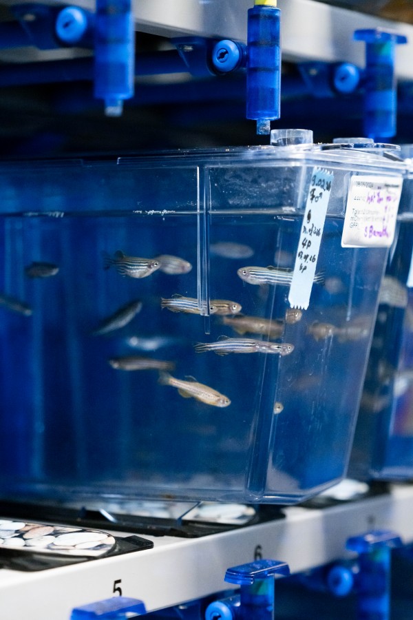
Small fish, big insights
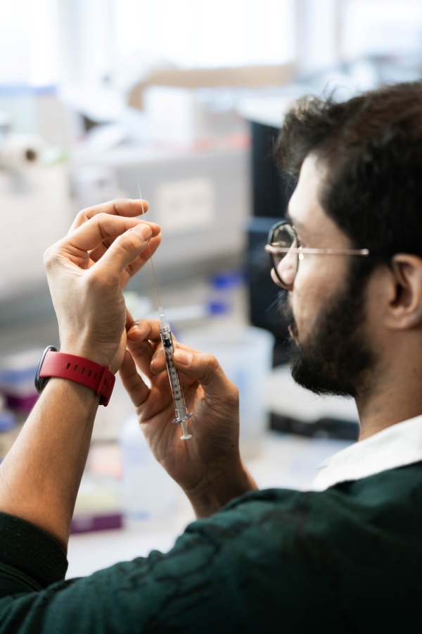
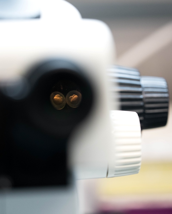
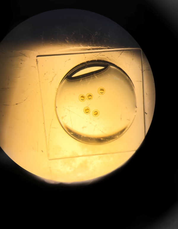
With a steady hand, Suyash Naik attaches a fine needle tip to a syringe and carefully maneuvers it into a petri dish. Drawing up a drop of water, he transfers it into a smaller dish that he places precisely under a microscope. Adjusting the focus, he sees five tiny spheres floating inside—zebrafish embryos.
“One major reason why we use zebrafish in basic research is that, unlike the commonly used fruit flyDrosophila, they are vertebrates,” explains Naik.
“They have a spinal cord similar to humans. Although their development differs greatly from that of mammals, they share many biological and evolutionary traits that help us connect the dots between species.”
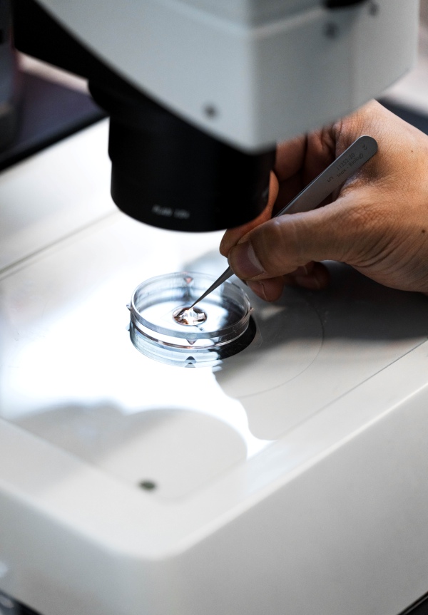
Another advantage is that zebrafish embryos are transparent and develop outside the mother’s body. That means they can be studied from the very moment of fertilization. The embryos under Naik’s microscope are only about one and a half hours old and are currently dividing—a stage known as the “cleavage period.”
Under careful observation, two tiny bubbles are clearly visible at the top of each sphere. These are the first cells to divide. Over the next few hours, they will become four, then eight, and so on—eventually forming a fully functional zebrafish.
How do cells move inside an embryo?
At this early stage, the embryo is still too young for Naik’s research. He is interested in a later phase—gastrulation—which in zebrafish typically takes place between five and ten hours after fertilization. During this period, a newly formed sheet of cells moves in a coordinated wave across the yolk, a process known as epiboly.
Driven by forces within a thin network of yolk cells—the yolk syncytial layer—this cellular sheet spreads down and around the yolk until it completely encloses the embryo in a protective layer. Inside, the first body layers—the germ layers—begin to take shape.
“You can think of it like pulling a cap down over your head until it covers your face,” Naik explains. “Except this cap never runs out of material—the cell layer that covers the embryo is amazingly elastic.”
The entire process happens quickly, yet it can still be observed in detail, making zebrafish embryos an ideal model for studying large-scale, coordinated cell movements and the forces that drive them.
Keratin – the softener
Keratins are filamentous proteins specific to epithelial tissues. There are many types of keratins, but the ones most familiar to us are found in our skin and hair, which give them strength. Many shampoos even boast of containing it.
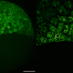
Inside embryonic cells, keratin forms filaments that look like tiny, curled noodles. Together with actin and myosin filaments, they make up the cytoskeleton—the internal frame of the cell.
As epiboly begins, keratin activity ramps up and continues to increase throughout the process. But exactly what role it plays had remained unclear—until now.

Keratin – the connector
This question became the focus of Naik’s PhD project. Using the gene-editing tool CRISPR-Cas9, he deleted the keratin genes in zebrafish embryos. When keratin was removed, epiboly slowed dramatically. Eventually, the entire cell layer collapsed.
Interestingly, the moving tissue became softer without keratin. That may sound counterintuitive; after all, with a rubber band, you’d expect that the softer it is, the easier it stretches. Here, the opposite was true.
Naik also observed that the cells within the tissue lost their proper alignment. The mechanical forces from the yolk syncytial layer could no longer be transmitted through the tissue, nor could the force from the yolk syncytial layer adapt to the movement.

What this means for the future
These results suggest that keratin acts as a critical connector for force transmission during epiboly. The study highlights just how versatile filament networks like keratin can be, serving as links between different structural systems within a tissue.
A deeper understanding of these interactions could reveal much about how cells coordinate movement during processes like wound healing or tissue regeneration and shed light on diseases such as Epidermolysis bullosa caused by keratin mutations, which can make tissues fragile, blistered, or prone to tearing. Understanding these interactions better would help us develop better skin-related interventions.

Publication:
Naik et al. 2026. Keratins coordinate tissue spreading by balancing spreading forces with tissue material properties. Nature Communications. DOI: 10.1038/s41467-026-72366-z
Funding information:
This project was supported by funding from the European Union’s Horizon 2020 research and innovation programme (grant agreement No.665385) and by the Austrian Science Fund (FWF) under projects PAT5044023 and W1250.
Information on animal studies:
To better understand fundamental processes, for example, in the fields of neuroscience, immunology, or genetics, the use of animals in research is indispensable. No other methods, such as in silico models, can serve as an alternative. The animals are raised, kept, and treated according to strict regulations.
