October 7, 2019
IST Austria inaugurates new cryo-electron microscopes
Austria’s largest and most advanced cryo-electron microscopy facility installed – Symposium on October 18 highlights developments in cutting-edge cryo technology
With three new state-of-the-art cryo-electron microscopes, IST Austria holds the largest and most advanced cryo-electron microscopy (cryo-EM) facility currently being operated in Austria. To inaugurate the new equipment, IST Austria hosts a cryo-EM symposium on October 18, 2019. The one-day event will highlight innovative research involving cryo-EM technology, ranging from the latest developments in the field to their application and general biological questions.
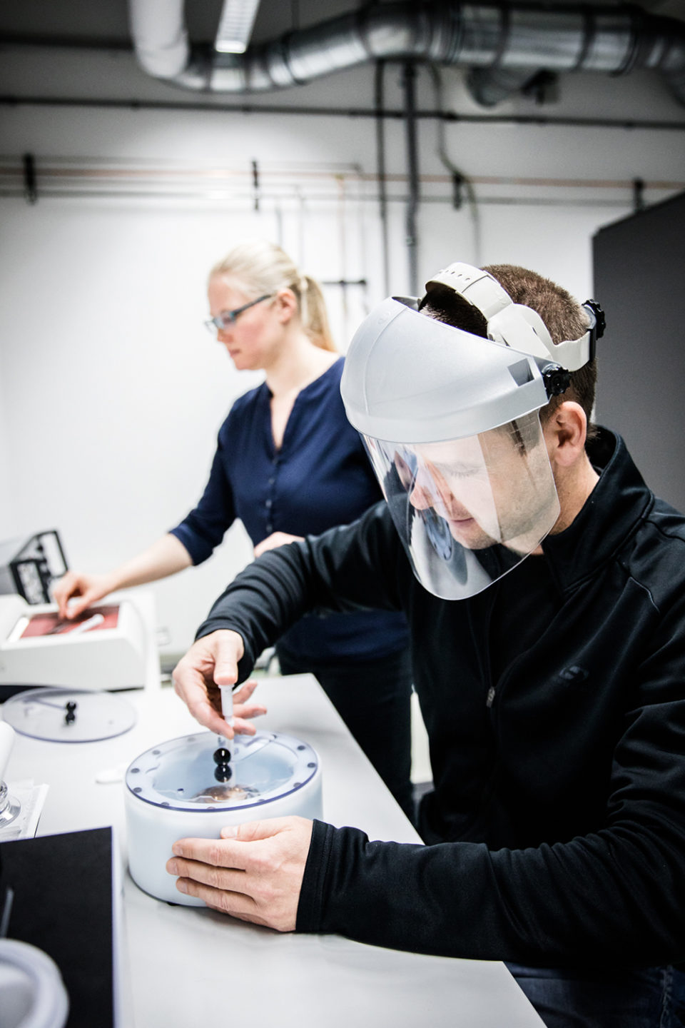
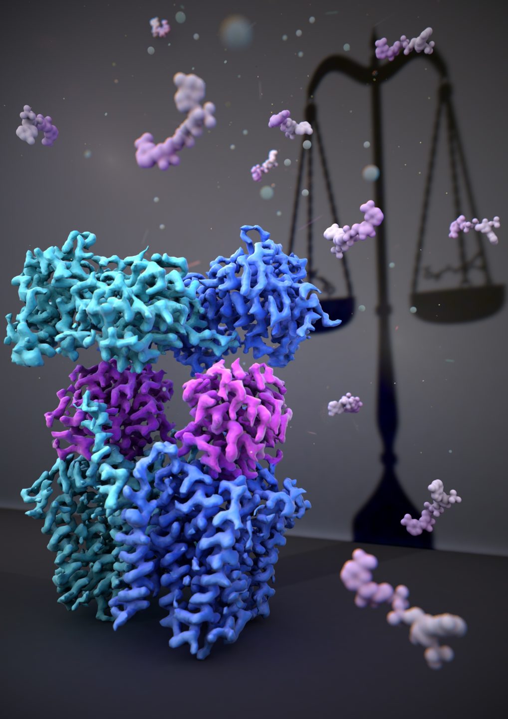
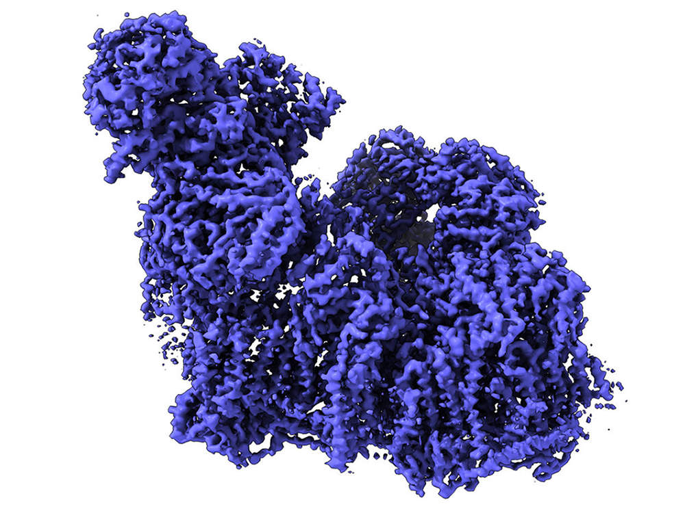
IST Austria has recently purchased and installed three state-of-the-art cryo-electron microscopes as part of the established and centrally organized Electron Microscopy Facility of the Institute. Cryo-EM is a cutting-edge technique, where biological samples such as proteins can be observed in their natural state and at near-atomic scales, rendering this method indispensable in structural biology. At IST Austria, cryo-EM is particularly being used by the research groups of three professors: Carrie Bernecky, Leonid Sazanov and Florian Schur. Significant research results have already been produced using the machines, including a paper in the journal Nature which was recently published by the group of Leonid Sazanov – the structural and functional analysis of the metabolic enzyme transhydrogenase.
Professor Leonid Sazanov on the advantages of the cryo-EM technology: “Classical imaging methods such as X-ray crystallography have failed to give a detailed look into the structure of some of the complex and flexible macromolecules. With the help of cryo-EM we can now look at even single protein domains at such high a resolution that we can draw conclusions about enzyme mechanics and functionality.”
Cryo-EM Symposium: October 18, 2019, at IST Austria, Klosterneuburg
With the “Cryo-EM Symposium” the new equipment will officially be inaugurated. Seven international experts will cover new developments in cryo-EM methodology as well as the subjects of cryo-EM single particle analysis and cryo-electron tomography. Besides scientific talks, the symposium offers a poster session and an optional tour of the new cryo-EM facility at IST Austria.
Speakers:
Pieter Abrahams, University of Basel, Switzerland
Peter Brzezinski, Stockholm University, Sweden
Gaia Pigino, Max Planck Institute of Molecular Cell Biology and Genetics, Germany
Jürgen Plitzko, Max Planck Institute of Biochemistry, Germany
Henning Stahlberg, University of Basel, Switzerland
Sharon Wolf, Weizmann Institute of Science, Israel
Peijun Zhang, University of Oxford, UK
Program details: https://cryo-em.ist.ac.at
The event is supported by Thermo Fisher Scientific and MiTeGen.
Background information
The new cryo-electron microscopies at the Electron Microscopy Facility of IST Austria
IST Austria’s cryo-EM facility includes the following machines:
- “300kV Cryo-TEM Titan Krios G3i”: a high-end 300kV transmission electron microscope for automated data acquisition for single-particle analysis and cryo-electron tomography
- “200kV Cryo-TEM Glacios”: a mid-range 200kV transmission electron microscope for automated sample screening and data acquisition for single-particle analysis and cryo-electron tomography
- “Leica cryo-CLEM”: a dedicated cryo-fluorescence microscope, designed for identification and targeting of fluorescently tagged structures in vitrified samples
- “Cryo-FIB/SEM Aquilos”: a system for the preparation of frozen, thin lamella from biological specimens for high-resolution tomographic imaging in a cryo-transmission electron microscope
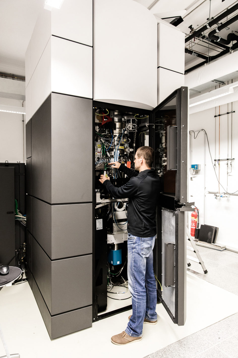
The new machines are part of the established Electron Microscopy Facility, one of eight individual Scientific Service Units (SSUs), i.e. facilities to pool scientific infrastructure used by more than one research group. Like all SSUs, the Electron Microscopy Facility is continuously working to optimize their equipment to cover the broadest possible range of applications. “With all three machines in full operation, we are able to do single-particle analysis, which is necessary to study proteins and viruses, as well as cryo-electron tomography, which enables the study of macromolecular complexes in their native cellular environment,” states Ludek Lovicar, manager of the Electron Microscopy Facility.
Cryo-electron microscopy (cryo-EM)
Cryo-electron microscopy is a cutting-edge technique that has led to a series of breakthrough discoveries in biology in the last years. While the development of the method began in the 1970s, recent advances in detector technology and software algorithms have allowed for the determination of biomolecular structures at near-atomic resolution. In 2017 the Nobel Prize in Chemistry was awarded jointly to Jacques Dubochet, Joachim Frank and Richard Henderson “for developing cryo-electron microscopy for the high-resolution structure determination of biomolecules in solution”. Today, cryo-EM has become an indispensable research tool that enables scientists to observe biological samples such as proteins in their natural state and at near-atomic scales.